

Echocardiography.
RAPID ACCESS to ESSENTIAL CARDIAC IMAGING.
An echocardiogram or simply ‘echo’, is a non-invasive type of heart scan that can obtain a large amount of information on heart function, muscle thickness, chamber size, valve function (narrowing or leak) and an indirect assessment of pressures (within the heart and lungs). As the name implies, echocardiography utilises high frequency sound waves called ultrasound, to generate images.
The strength of echocardiography is in its versatility - it is non-invasive, harmless and painless, capable of accurately detecting and monitoring a wide variety of cardiac conditions. Echocardiography has replaced invasive assessment for many conditions over the years.
Common indications for an echo are:
Assessment of left ventricular (LV) systolic and diastolic function (main heart pump). The LV ‘ejection fraction’ (LVEF) is often used as a cut-off for certain drugs and interventions. Normal ejection fraction is usually ≥50%. Reduced ejection fraction can be a feature of Heart Failure (HFrEF - heart failure with reduced ejection fraction), and cause symptoms such as breathlessness, fatigue and fluid build up. Diastolic dysfunction can also cause heart failure (HFpEF - heart failure with preserved ejection fraction).
Assessment of right ventricular (RV) function e.g. in rare cardiac conditions, lung conditions.
Establishing risk of arrhythmias and guiding their management.
Assessment of valve disorders such as Aortic Stenosis and Mitral Regurgitation, indications for valve intervention (e.g. percutaneous and surgical techniques).
Assessment following a heart attack.
Assessment of inherited and rare heart muscle disorders or ‘cardiomyopathies’ e.g. Dilated Cardiomyopathy, Hypertrophic Cardiomyopathy.
Assessment of Congenital Heart Disease e.g. Bicuspid Aortic Valve, atrial septal defects (ADS), ventricular septal defects (VSD) or more complex congenital heart disease in adults that has been operated on in childhood e.g. Tetralogy of Fallot, a Fontan circuit, transposition of the great arteries.
Assessing fitness for organ transplant, fitness for non-cardiac surgery (e.g. a new hip, abdominal or vascular surgery), fitness to drive, HGV licencing, monitoring effect of chemotherapy and medications that can be toxic to the heart, assessment in professional athletes.
OUR PROMISE
We pride ourselves in being able to facilitate echo rapidly. Our average waiting time is only 2 weeks.
Public sector waiting times are exceptionally long (>30 weeks in some health boards), reflecting real service and resource pressures. However, for debilitating symptoms such as chest pain, breathlessness and palpitations, this is not acceptable. Likewise, certain conditions need rapid assessment. For example, sudden loss of consciousness where a structural heart abnormality is suspected needs prompt assessment. Long delays to assessing certain rhythm conditions such as Atrial Fibrillation, can risk leaving things ‘too late’ past the ideal intervention window. Severe valve disorders can carry a high mortality; severe symptomatic aortic stenosis for example has a life expectancy of 1-2 years. Prompt assessment provides clarity over diagnosis, treatment, peace of mind, resolves questions from insurers as well as employers.
Our Sonographers are British Society of Echocardiography (BSE) accredited experts with vast experience in performing echo, provide training and take part in clinical research.
We will only issue full, detailed echo reports to BSE standard. This includes all the technical measurements required to make concrete decisions about treatments, further assessments and interventions (e.g. what tablets to recommend, device related queries, surgical cut-offs, surveillance cycles). We do not simply describe the key findings qualitatively in your clinical correspondence. Performing an echo without comprehensive assessment to BSE standard and record of technical measurements/parameters, can result in harm and have serious medicolegal implications.
Your echo report is produced the same day of scanning and a digital copy is provided to you and your referring Specialist.
Your GP or Specialist can request an echo directly via our Referrals page.
If you think you require an echo, you will need to Contact Us first to have a consultation with one of our Cardiologists.
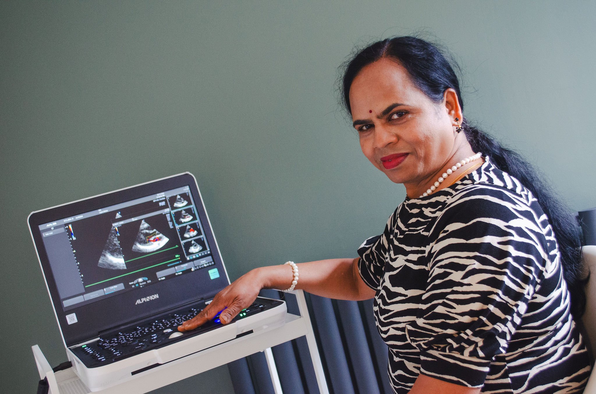
MEET YOUR SONOGRAPHER

Mrs. Janaki Srinivasan
BSc, DMLT, Dip, BSE
Chief Cardiac Sonographer
Mrs. Srinivasan is a Chief Cardiac Physiologist specialising in Echocardiography. Her career in the NHS began in 2005 as a Chief Cardiac Physiologist, obtaining membership of the British Society of Echocardiography in 2008. She continues to offer her services in both NHS and Private Medical capacities.
Mrs. Srinivasan is highly experienced in all aspects of echocardiography required for the accurate diagnosis and ongoing assessment of a range of cardiac conditions. This includes strain analysis, stress echocardiography, transeosophageal echocardiography (TOE) as well as congenital heart disease.
In parallel to her clinical activity Mrs Srinivasan is actively involved in a number of research studies that require both standard and experimental echocardiographic techniques. She is also an experienced educator in the field.